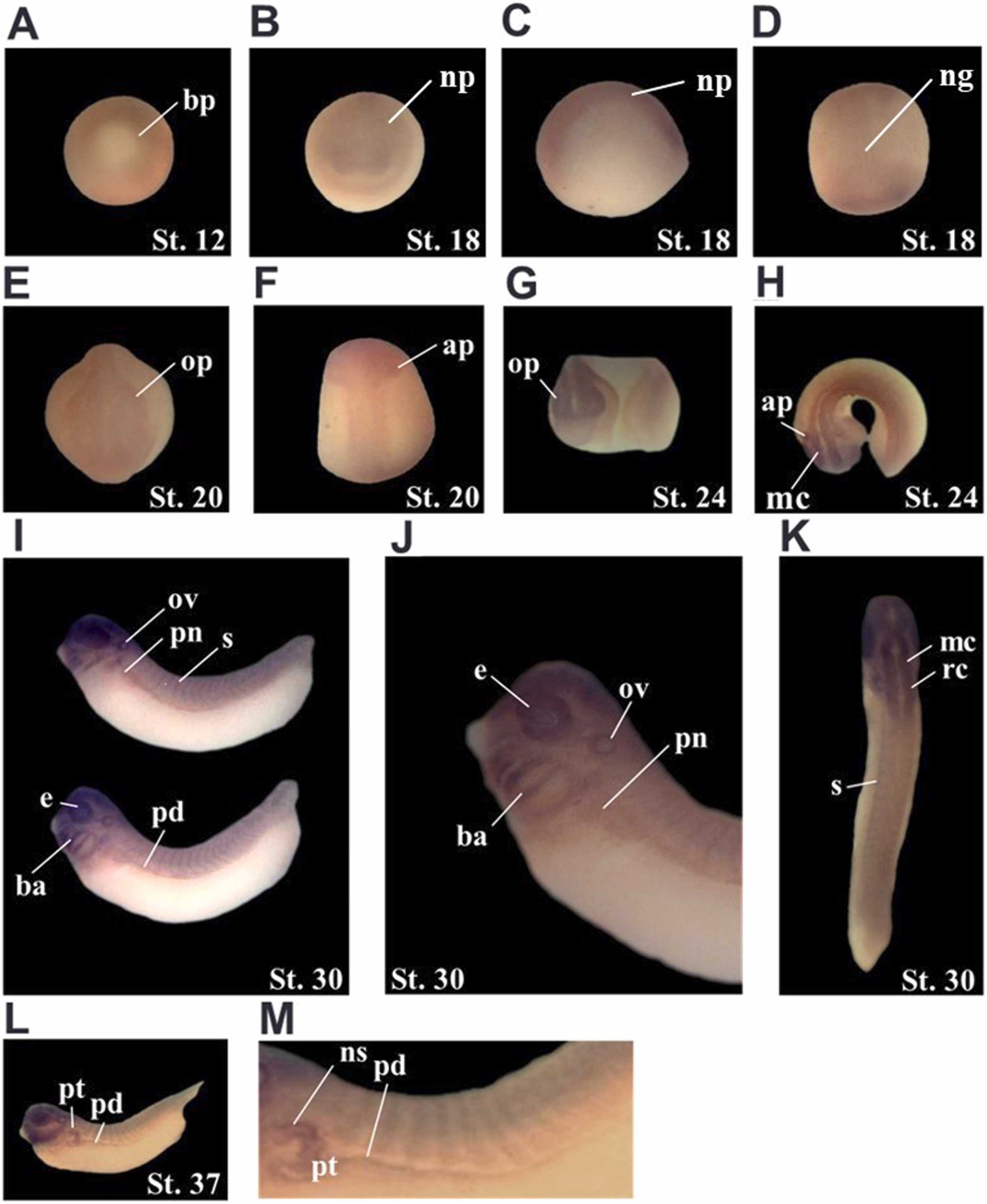
Fig. 2. Spatial expression patterns of XPofut1. Whole-mount in situ hybridization was performed with embryos of indicated stages. Photographs of representative embryos are shown. (A) Stage 12, vegetal view, dorsal side is toward the top. (B-D) Stage 18, anterior view, dorsal side is toward the top (B), lateral view, dorsal side is toward the top (C), and dorsal view, anterior side is toward the top (D). (E and F) Stage 20, anterior view, dorsal side is toward the top (E) and dorsal view, anterior side is toward the top (F). (G and H) Stage 24, lateral view (G), and dorsal view (H). (I-K) Stage 30, lateral view (I), higher magnification of anterior region (J), and dorsal view (K). (L and M) Stage 37, lateral view (L) and higher magnification of dorsal region (M). ap, auditory placode; ba, branchial arch; bp, blastopore; e, eye; mc, mesencephalon, ng, neural groove; np, neural plate; ns, nephrostome; op, optic placode; ov, optic vesicle; pd, pronephric duct; pn, pronephros; pt, pronephric tubule; rc, rhombencephalon; s, somite.
Image published in: Kim YJ et al. (2025)
© 2025 The Author(s). Creative Commons Attribution license
| Gene | Synonyms | Species | Stage(s) | Tissue |
|---|---|---|---|---|
| pofut1.L | fut12, o-fuc-t, o-fuct-1, o-fut, XPofut1 | X. laevis | Throughout NF stage 12 | marginal zone (sensu neural) |
| pofut1.L | fut12, o-fuc-t, o-fuct-1, o-fut, XPofut1 | X. laevis | Sometime during NF stage 18 to NF stage 20 | neural plate optic field anterior neural tube cranial neural crest chordal neural plate |
| pofut1.L | fut12, o-fuc-t, o-fuct-1, o-fut, XPofut1 | X. laevis | Throughout NF stage 24 | eye primordium neural tube midbrain otic placode |
| pofut1.L | fut12, o-fuc-t, o-fuct-1, o-fut, XPofut1 | X. laevis | Throughout NF stage 29 and 30 | otic vesicle pharyngeal arch mandibular arch hyoid arch branchial arch somite eye pronephric kidney pronephric duct brain forebrain midbrain hindbrain |
| pofut1.L | fut12, o-fuc-t, o-fuct-1, o-fut, XPofut1 | X. laevis | Throughout NF stage 37 and 38 | pronephric kidney pronephric nephrostome pronephric duct pronephric tubule eye brain pharyngeal region somite |
Image source: Published
Permanent Image Page
Printer Friendly View
XB-IMG-204400