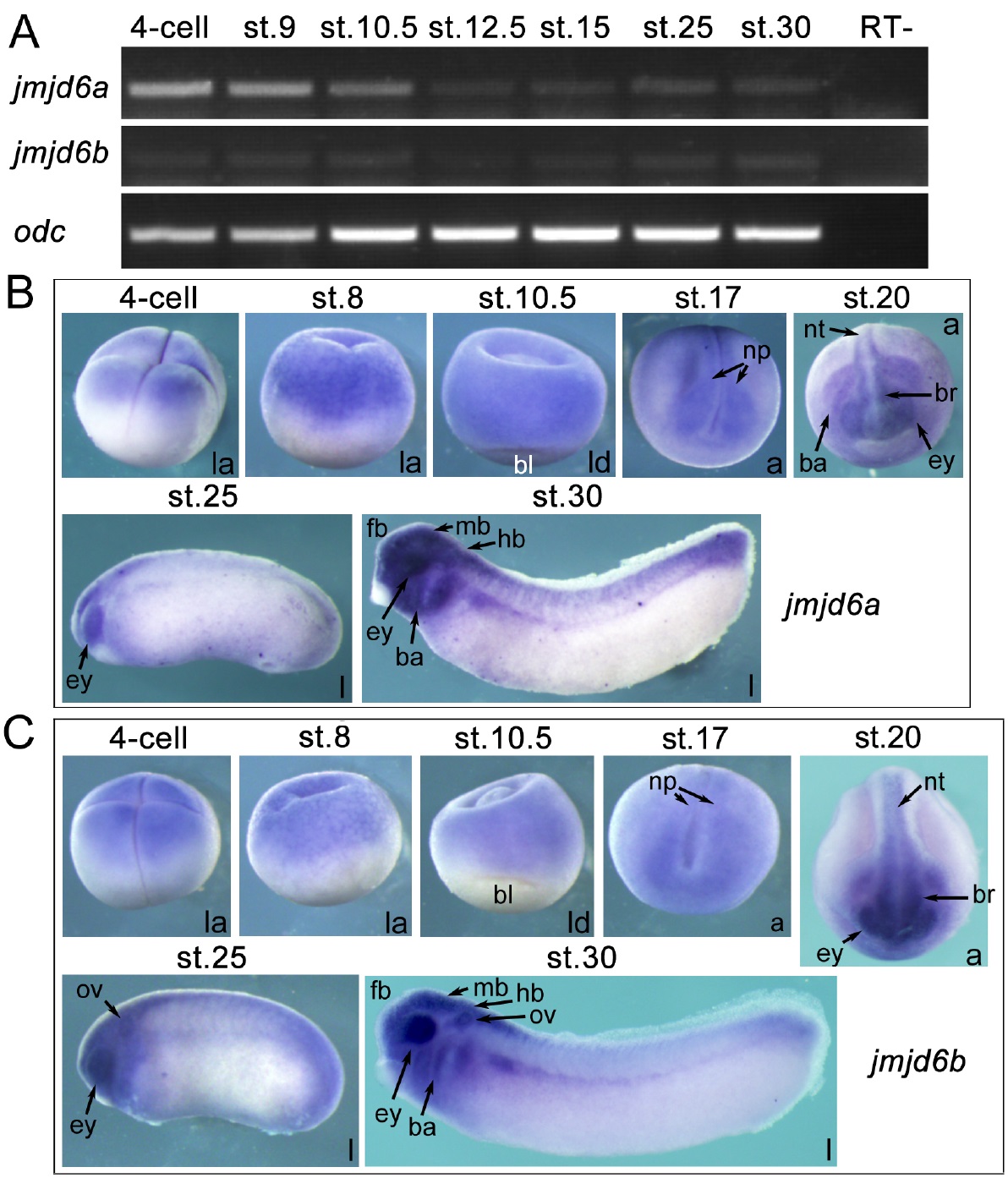
FIGURE 5. The spatio-temporal expression patterns of jmjd6a and jmjd6b during the embryogenesis of Xenopus laevis. A, temporal expression of jmjd6a (Accession number: NM_001092479) and jmjd6b (Accession number: NM_001087045) in different stages of embryos detected with RT-PCR. Expression of odc was used as a loading control. RT-: transcription without reverse transcriptase. B and C, spatial expression patterns of jmjd6a (B) and jmjd6b (C) detected with whole mount in situ hybridization. Embryo stages are indicated at the top of each panel. a: anterior view, with the dorsal at the top of the panel; ba: branchial arch; bl: blastopore lip; br: brain; ey: eye; fb: forebrain; hb: hindbrain; l: lateral view, with the anterior to the left; la: lateral view, with animal pole at the top; ld: lateral-dorsal view, with the animal pole at the top; mb: midbrain; np: neural plate; nt: neural tube; ov: otic vesicle.
Image published in: Zhang X et al. (2015)
Copyright © 2015. Image reproduced with permission of the Publisher and the copyright holder. This is an Open Access article distributed under the terms of the Creative Commons Attribution License.
| Gene | Synonyms | Species | Stage(s) | Tissue |
|---|---|---|---|---|
| jmjd6.L | jmjd6-a, jmjd6a, jmjd6-b, psr, ptdsr, ptdsr1 | X. laevis | Sometime during NF stage 17 to NF stage 20 | neural plate optic vesicle cranial neural crest branchial crest neural tube posterior neural tube anterior neural tube |
| jmjd6.L | jmjd6-a, jmjd6a, jmjd6-b, psr, ptdsr, ptdsr1 | X. laevis | Throughout NF stage 25 | central nervous system spinal cord brain hindbrain |
| jmjd6.L | jmjd6-a, jmjd6a, jmjd6-b, psr, ptdsr, ptdsr1 | X. laevis | Throughout NF stage 29 and 30 | eye pharyngeal arch hyoid arch branchial arch pronephric mesenchyme central nervous system spinal cord brain forebrain midbrain hindbrain |
| jmjd6.L | jmjd6-a, jmjd6a, jmjd6-b, psr, ptdsr, ptdsr1 | X. laevis | Throughout NF stage 3 (4-cell) | animal pole animal blastomere |
| jmjd6.L | jmjd6-a, jmjd6a, jmjd6-b, psr, ptdsr, ptdsr1 | X. laevis | Sometime during NF stage 8 to NF stage 10.5 | animal hemisphere ectoderm |
Image source: Published
Permanent Image Page
Printer Friendly View
XB-IMG-145589