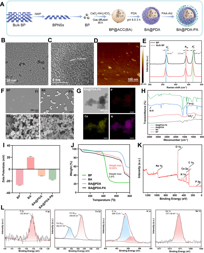
Fig. 1. Formation and characterization. (A) Diagram of the synthesis of BA@PDA-PA. (B) TEM image of BP. (C) HRTEM image of BP. (D) AFM image of BP. (E) Raman spectra of BP and bulk BP. (F) SEM images of BP, BA, BA@PDA and BA@PDA-PA. (G) TEM and mapping images of BA@PDA-PA. (H) FTIR spectra of BP, BA, BA@PDA and BA@PDA-PA. (I) Zeta potentials of BP, BA, BA@PDA and BA@PDA-PA. (J) TGA curves of BP, BA, BA@PDA and BA@PDA-PA. (K) XPS survey spectrum of BA@PDA-PA. (L) High-resolution XPS spectra of P 2p, Ca 2p, N 1 s and Na 1 s in BA@PDA-PA
Image published in: Shen X et al. (2026)
Image downloaded from an Open Access article in PubMed Central. © The Author(s) 2026
Permanent Image Page
Printer Friendly View
ECB-IMG-208666