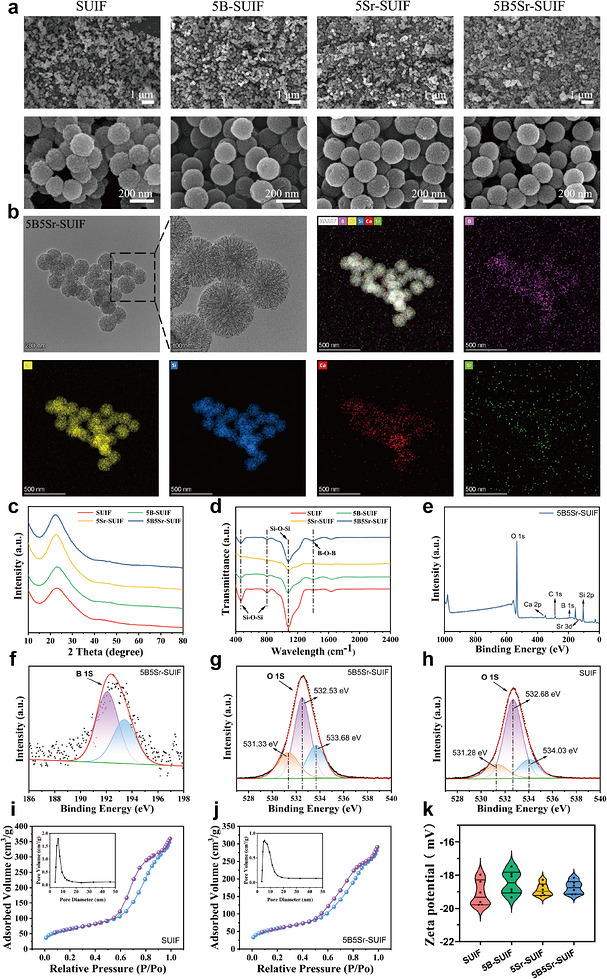
FIGURE 1. Morphology and physicochemical characterization of SUIF‐based nanoparticles. (a) SEM micrographs of SUIF, 5B‐SUIF, 5Sr‐SUIF, and 5B5Sr‐SUIF. (b) TEM image and corresponding elemental mapping of 5B5Sr‐SUIF. (c) XRD patterns of SUIF, 5B‐SUIF, 5Sr‐SUIF, and 5B5Sr‐SUIF. (d) FTIR spectra of SUIF, 5B‐SUIF, 5Sr‐SUIF, and 5B5Sr‐SUIF; (e) XPS survey spectrum of 5B5Sr‐SUIF. (f) high‐resolution B 1s XPS spectrum of 5B5Sr‐SUIF; (g) high‐resolution O 1s XPS spectrum of 5B5Sr‐SUIF. (h) High‐resolution O 1s XPS spectrum of SUIF. (i) N2 adsorption–desorption isotherm of SUIF; (j) N2 adsorption–desorption isotherm of 5B5Sr‐SUIF. (k) Zeta potential profiles of SUIF, 5B‐SUIF, 5Sr‐SUIF, and 5B5Sr‐SUIF.
Image published in: Lu Y et al. (2026)
Image downloaded from an Open Access article in PubMed Central. © 2026 The Author(s). Endocrinology, Diabetes & Metabolism published by John Wiley & Sons Ltd.
Permanent Image Page
Printer Friendly View
ECB-IMG-208619